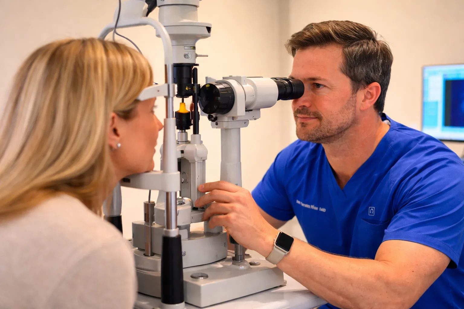
Dry eye is not diagnosed with a single test. We measure how long your tears last, how much there is, the condition of your meibomian glands, and whether the surface of the eye already shows damage. None of the tests hurts, and most do not even touch the eye. The results tell us what is causing your symptoms, and the cause determines the treatment.
Why measure
How is dry eye diagnosed?
Two people with the same complaints can have opposite causes. One produces too few tears; the other produces enough, but they evaporate because the meibomian glands are blocked. The symptom is the same, and the treatment is different.
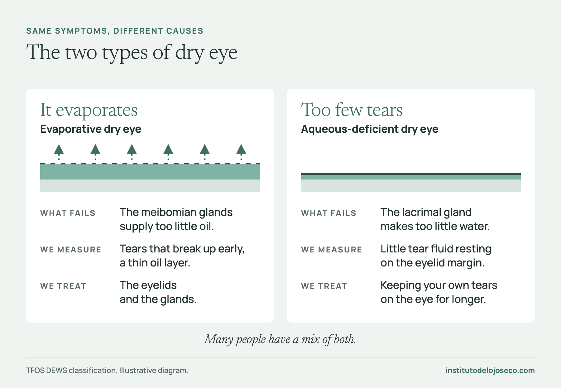
The international TFOS DEWS III criteria require two things for a diagnosis of dry eye disease: symptoms, and at least one measurable sign that the tears or the ocular surface have lost their balance, such as tears that break up too soon, tears with too much salt, or visible damage to the surface. The remaining tests serve to find the cause and to measure, at each follow-up, whether the treatment is working.
What the visit is like
All the tests are done at the institute, at Dante 6, Colonia Anzures, Mexico City.
- 01
Medical history and habits
Your symptoms, screen time, environment, contact lenses, medications, and previous surgeries, plus a twelve-question questionnaire (OSDI), which you can complete ahead of time with the dry eye test.
- 02
Imaging measurements
With no dyes and no contact: how long your tears last, how much there is, the condition of their oil layer, how red the eye is, and how your glands look. The salt concentration is measured with a tiny tear sample.
- 03
Examination with the office microscope
With the slit lamp, we examine the eyelid margins, the gland openings, and the eyelashes. Last comes staining of the cornea and conjunctiva; it goes at the end because the dye alters the tears and would skew the earlier measurements.
- 04
Your diagnosis and your plan
We show you your images, tell you which cause is dominant, and define the goal of treatment and its follow-up.
The tests
What each test answers
| Test | What it answers | How it is done |
|---|---|---|
| OSDI questionnaire | What you feel and how much it affects your day | Twelve questions |
| Tear breakup time (NIBUT) | How many seconds your tears last | Imaging, no contact |
| Tear meniscus | How much tear fluid there is in your eye | Imaging, no contact |
| Lipid layer | Whether the oil is enough to slow evaporation | Imaging, no contact |
| Meibography | Whether your glands are intact or have been lost | Imaging; the eyelid is turned over for a few seconds |
| Osmolarity | How concentrated in salts your tears are | A tiny tear sample |
| Redness | How much inflammation there is | Imaging, no contact |
| Fluorescein staining | Whether the surface of the eye already has damage | A drop of dye |
Tear tests
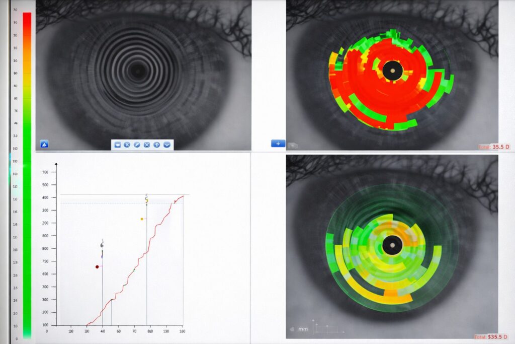
NIBUT: noninvasive tear breakup time
It measures how many seconds your tears stay stable after a blink, with no dyes and no contact, and it takes seconds. Tears that break up in less than 10 seconds are unstable: this is one of the signs that confirm the diagnosis.
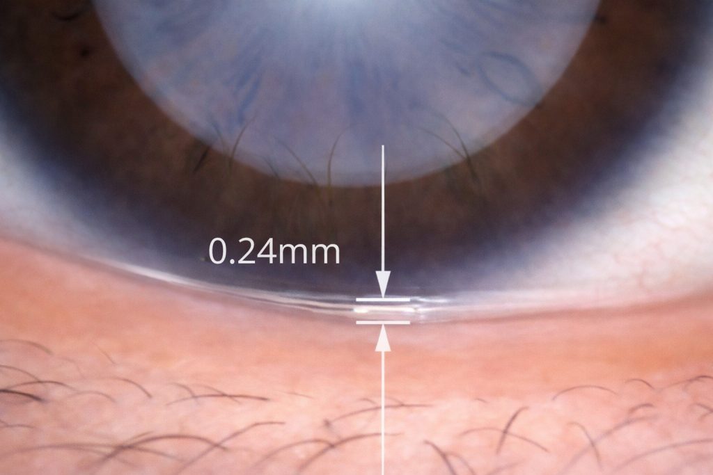
Tear meniscus height
An image, taken without contact, measures the strip of tears that collects along the edge of the lower eyelid. A low meniscus suggests that the lacrimal gland is producing too few tears, in other words, aqueous-deficient dry eye.
Lipid layer
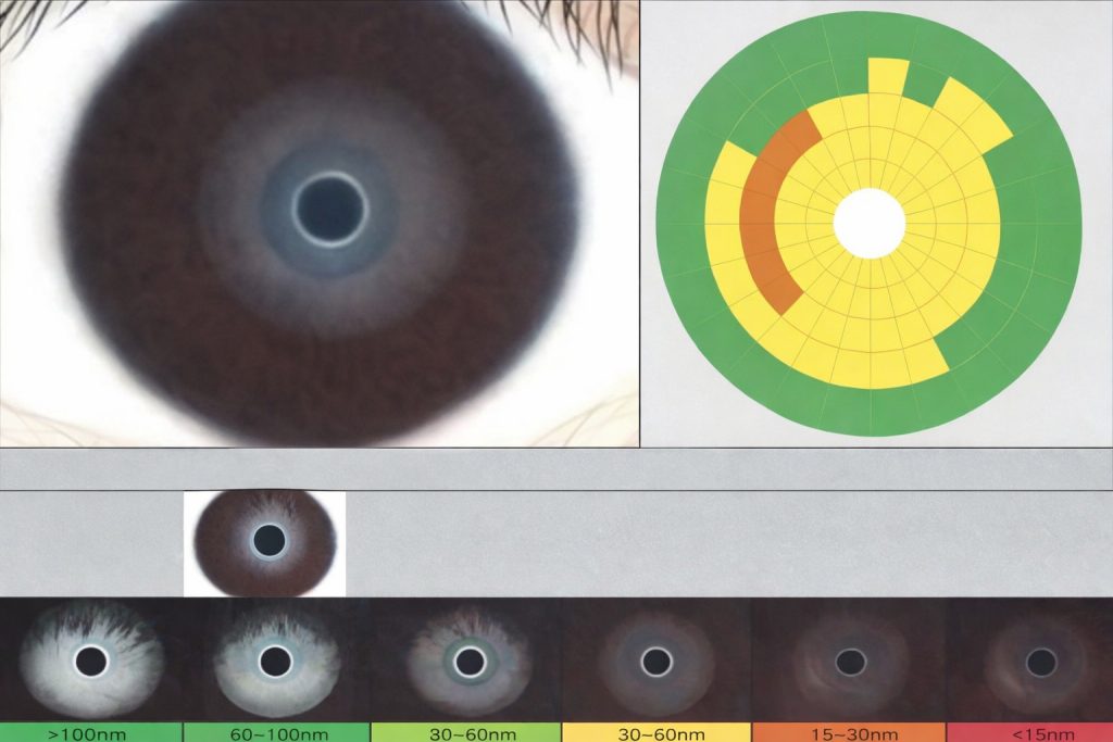
This test analyzes the thickness and distribution of the oily layer of the tears while you blink naturally; it takes one to two minutes. A thin layer usually points to meibomian gland dysfunction, because those glands are the ones that produce it.
Tear osmolarity
It measures the salt concentration of the tears. When tears are too concentrated, they damage the surface cells and feed inflammation. A large difference between the two eyes is also a sign of dry eye.
Meibography and eyelid tests
Meibography
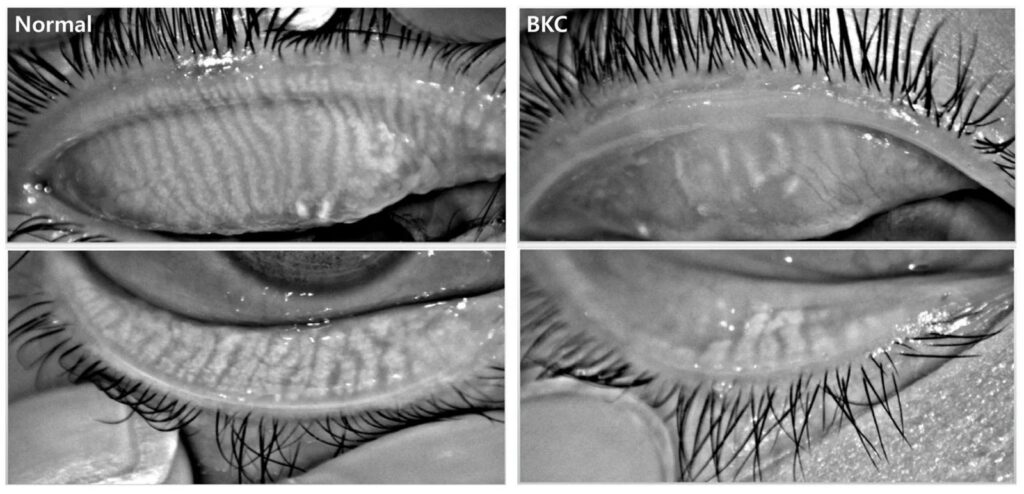
An infrared image of the glands inside the eyelid: it shows whether they are intact, shortened, or twisted, or whether they have already disappeared. To see them, the eyelid is gently turned over for a few seconds. Gland loss is usually irreversible, which is why it is worth detecting early.
Gland openings
With each blink, the glands release their oil through tiny openings along the eyelid margin. If the openings become plugged, the oil does not reach the tears and evaporative dry eye develops; if the blockage continues, the gland can atrophy. Seeing the condition of the openings guides the first step of treatment.
Eyelashes and eyelid margin
Inflammation of the eyelid margin (blepharitis) usually combines an overgrowth of bacteria, meibomian gland dysfunction and, in some cases, rosacea or Demodex, a mite that lives at the root of the eyelashes. With high-resolution images we look for crusting, redness, and cylindrical dandruff (collarettes) at the base of the lashes, the typical deposit left by Demodex.
Eyelid margin blood vessels
Tiny dilated vessels along the eyelid margin, called telangiectasias, feed inflammation and can point to dry eye associated with rosacea. Their presence is one of the reasons to recommend intense pulsed light (IPL).
Tests of the surface of the eye
Eye redness
An image of the white of the eye grades the redness. It is used to measure the intensity of the inflammation and compare it at each follow-up.
Corneal and conjunctival staining
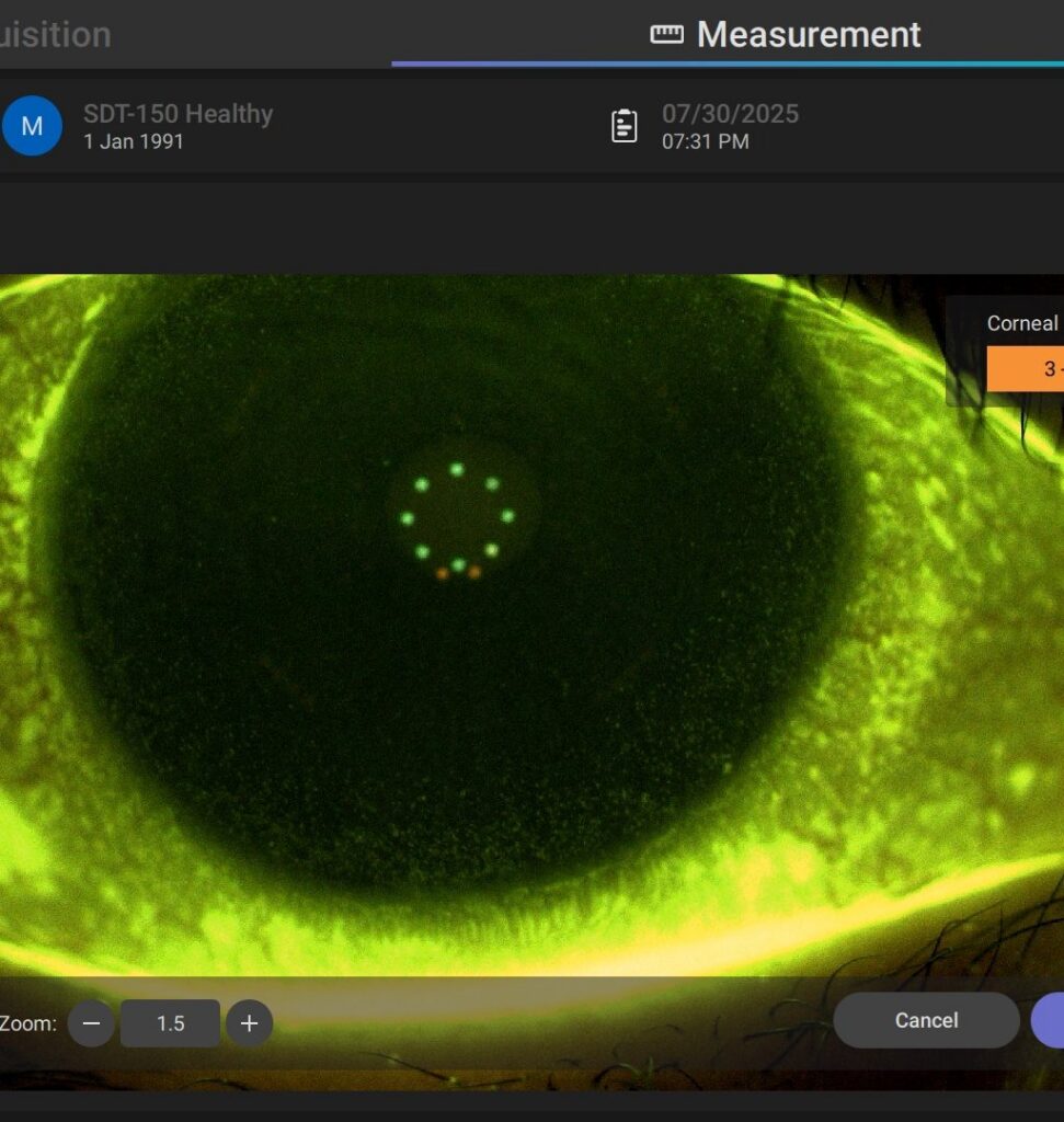
A drop of fluorescein, a safe dye, makes dry spots and surface damage visible under blue light. The pattern and intensity of the staining show how advanced the disease is, help identify the cause, and leave a clear baseline for evaluating how treatment is progressing.
What happens after the diagnosis?
With the results we name the dominant cause: blocked glands that let the tears evaporate, low tear production, inflammation of the eyelid margin, Demodex, or ocular rosacea. It is almost always a combination, and so is the plan: eyelid cleaning, infrared thermotherapy, meibomian gland expression, IPL, LLLT light therapy, punctal plugs, or medical treatment, depending on what we find.
The same measurements, repeated at follow-up visits, show whether the plan is working and when it should be adjusted.
Sources
- Wolffsohn JS, Benítez-del-Castillo JM, Loya-Garcia D et al., TFOS DEWS III: Diagnostic Methodology. American Journal of Ophthalmology, 2025. PubMed
Book your diagnostic visit
We measure your tear film, your glands, and your ocular surface and explain what is causing your symptoms.
Common questions
Frequently asked questions
Do the tests hurt?
No. Most are done with imaging, without touching the eye; for meibography the eyelid is turned over for a few seconds, which can be slightly uncomfortable but does not hurt.
Do I have to stop wearing my contact lenses beforehand?
Let us know when you book if you wear them: we will tell you whether you should stop wearing them a few days before. On the day of your appointment, come wearing your eyeglasses and without eye makeup. More in Book an assessment.
I already know I have dry eye. Why do I need a diagnosis?
To find out which type it is and what is causing it. Eye drops do not unclog a blocked gland or control Demodex blepharitis; without measurements, treatment is guesswork.
Is it useful if I’m having cataract or refractive surgery?
Yes. Unstable tears distort the corneal measurements and the intraocular lens calculation. Stabilizing the surface before surgery helps make those measurements reliable. More in Dry eye before and after eye surgery.